
Transformation
Replication of Clifford Carnicom's electromagnetic blood experiments

The basis for this article and experiment is the following paper:
https://carnicominstitute.org/blood-alterations-iii-transformation/
From that paper, I quote Clifford Carnicom:
”There is no possibility of avoiding the following statement, and it is declared at the onset.
Direct evidence indicates that the application of a low magnitude electrical current can or will transform a blood sample to the point that it is no longer recognizable as blood in any conventional sense, and the nature of the blood is then dominated by the presence of the cross-domain bacteria (CDB) (CI nomenclature) microbial life form and its manifestations.”
All my work is based on the decades of research done by Clifford Carnicom/CI. This should be the first place readers look for additional information.
This post will consist of images and video footage of the apparent transformation of the blood into a substance (likely synthetic tissue scaffolding) comprised mostly of CDBs (Cross Domain Bacteria - CI nomenclature for now ubiquitous synthetic biology) and their associated secreted protein(s) and other metabolic products.
The Carnicom Institute (CI) has decades of research on this highly engineered and pathogenic ‘microbe’; the following paper is just a more recent one out of many.
https://carnicominstitute.org/cross-domain-bacteria-cdb-protein-the-fallout-emerges/
I have repeated this experiment many times in many configurations.
This is transhumanism.
We will now turn attention to a more recent replication experiment I have performed, which illustrates this transformation in a rather vivid manner.
An image of the particular experimental fixture:
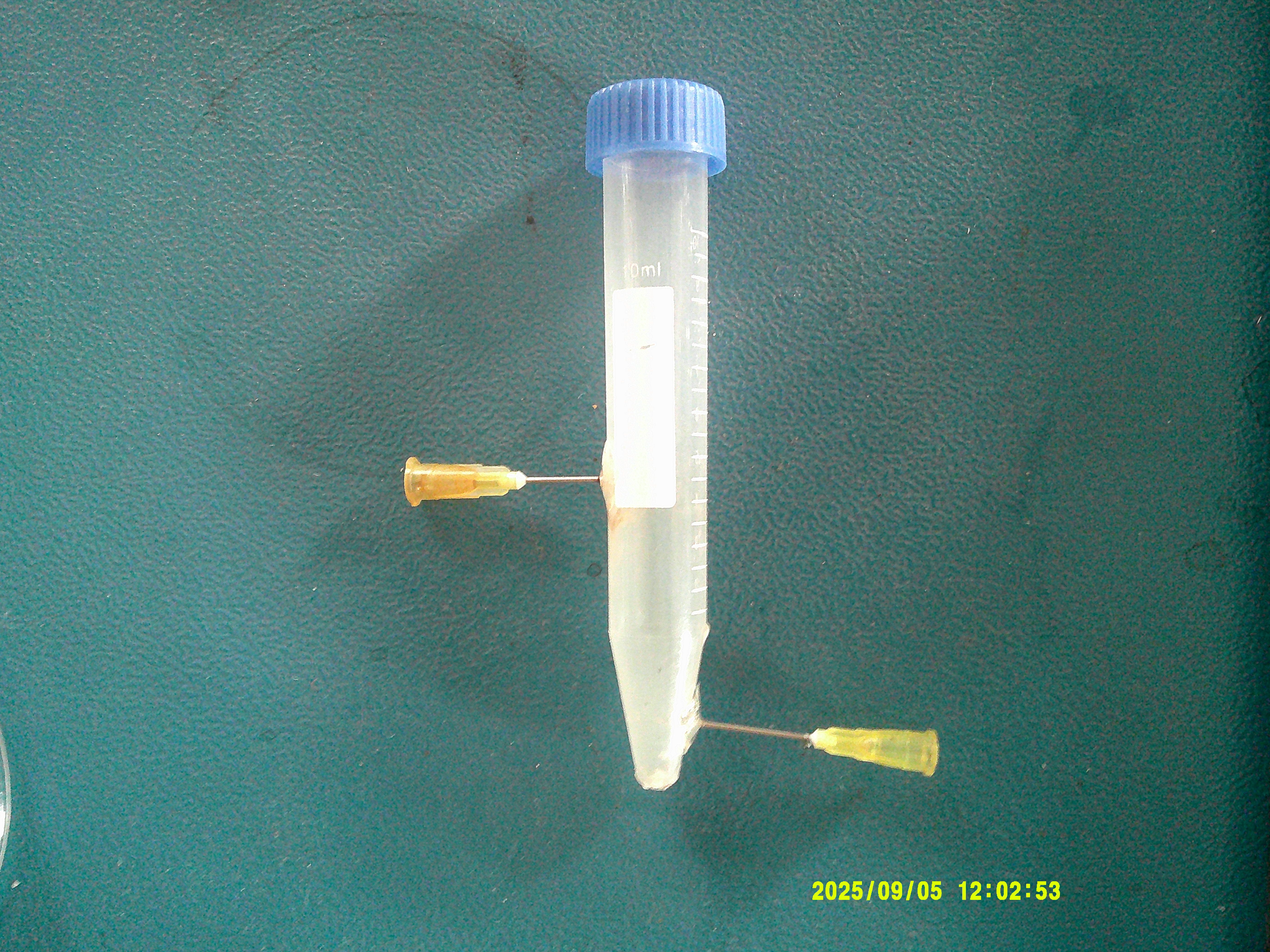
The tube has stainless steel needles inserted into the top and bottom. These serve as the electrodes, which are attached to the power supply via clips. This allows for an electrical circuit to be completed through the blood/water mixture.
A mixture of 10 drops of venous blood from a 29g insulin needle and approximately 6-7mL of distilled water was added to the tube:
Electrical energy supplied by a benchtop DC adjustable power supply.
There is nothing more to this experiment; it can easily be done with a single battery.
There are a number of ‘stages’ that the reaction proceeds through; I have documented these in some detail, and will share them in a future post.
For now, we shall skip to the ‘end result’ of this process (approximately 2 days after beginning); applied voltage 6VDC:
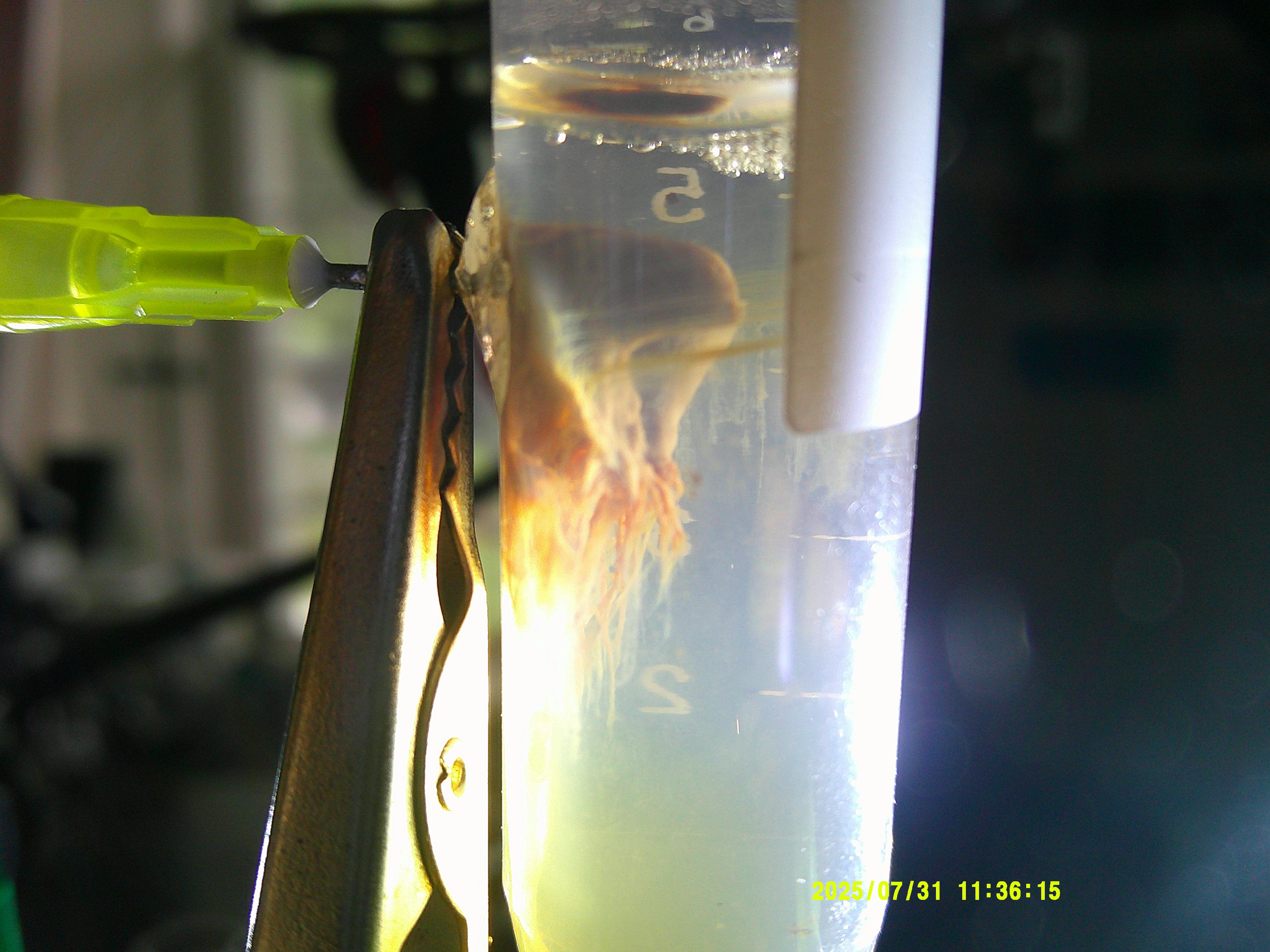
I do beg readers to keep foremost in mind that this was a simple, homogenous mixture of 10 drops of blood from a 29g insulin needle and 6mL of distilled water, and an applied voltage of 6VDC over 2 days.
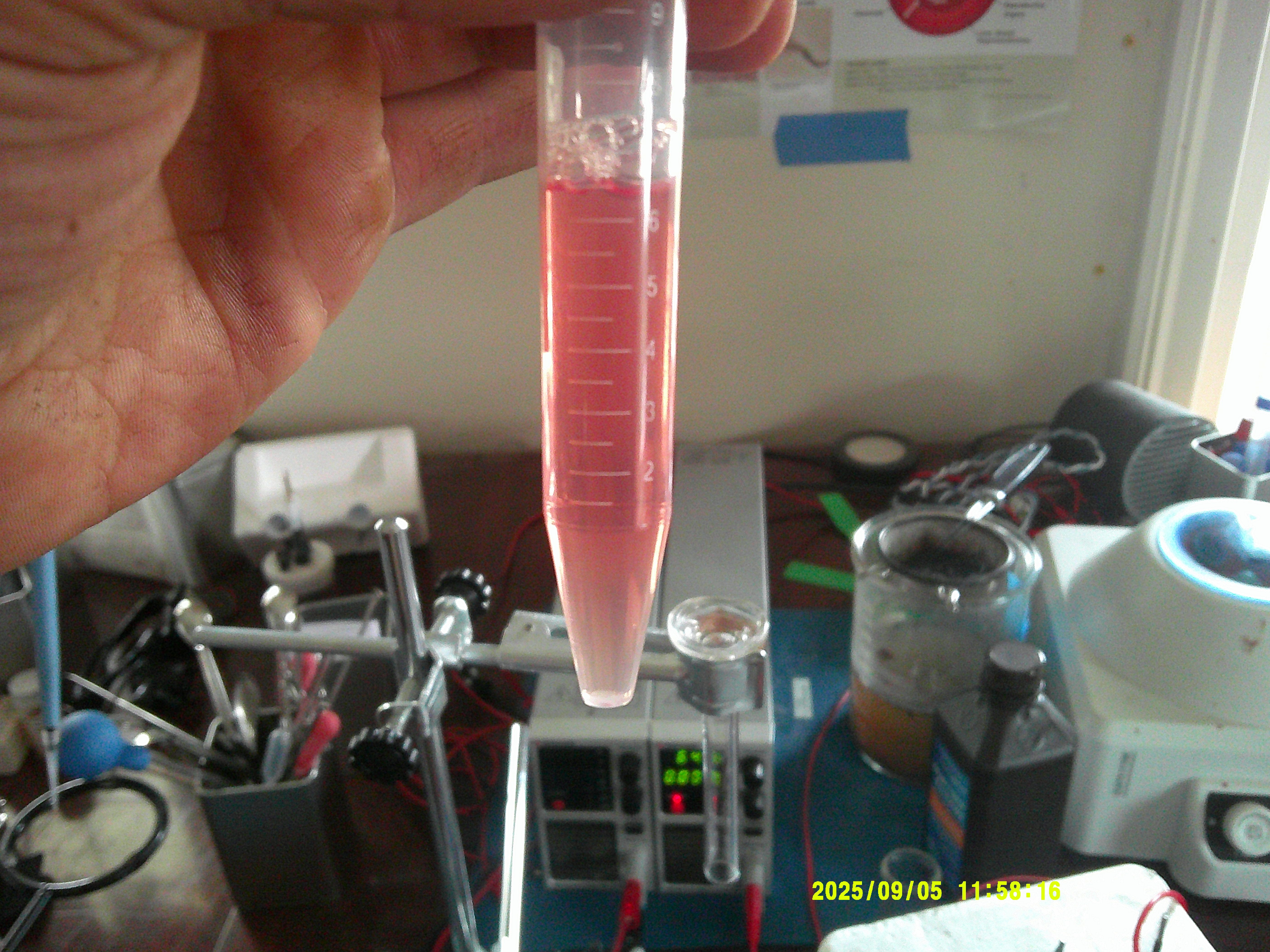
This is what 10 drops of blood in the tube looks like, for comparison:
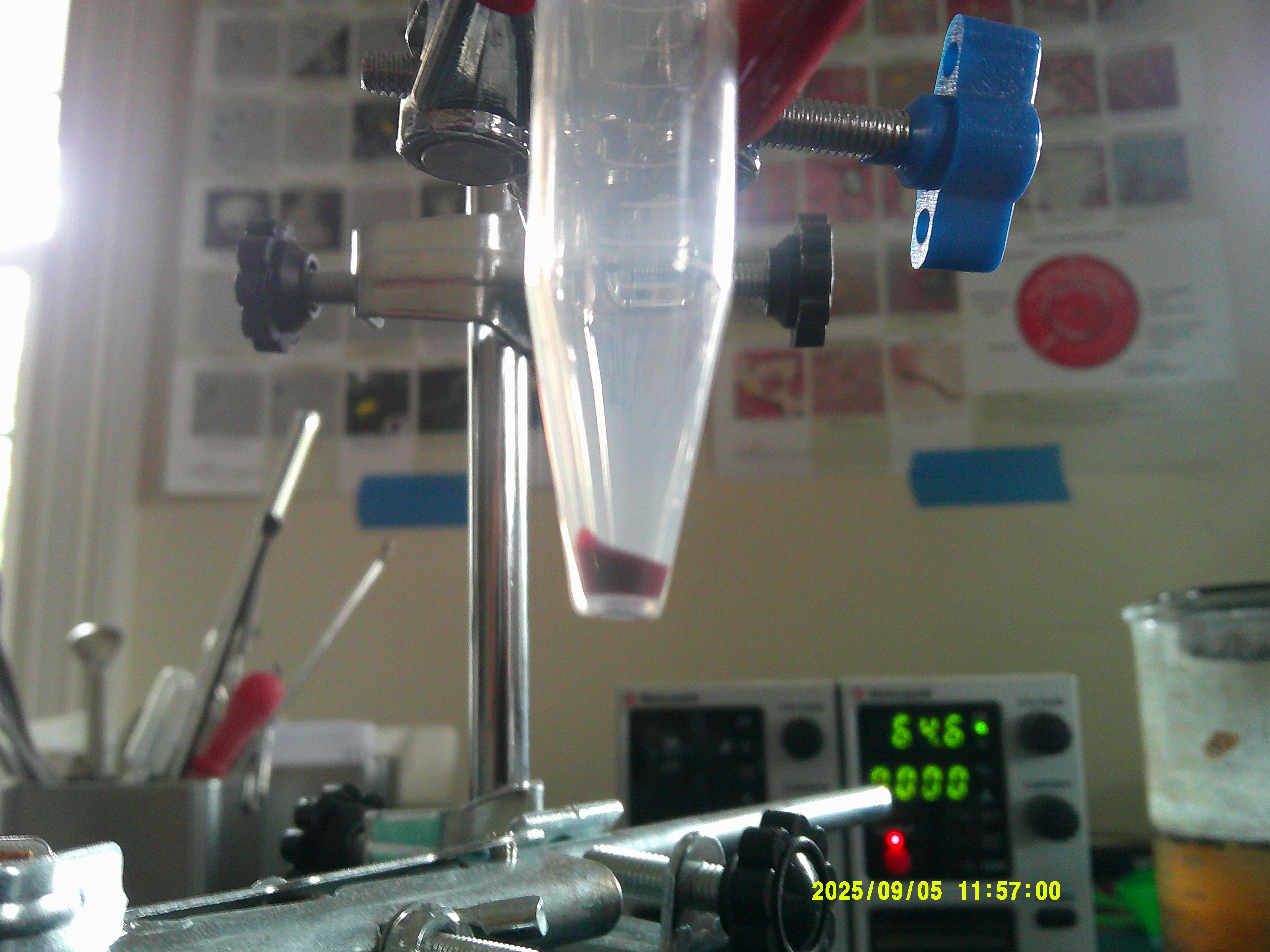
Further images of suspected synthetic tissue scaffolding.


As can be seen in one of the above images, it appears that the blood itself (or at least the coloring) remains at the bottom of the tube.
The filamentous material is highly extensive, and is affixed to the wall of the tube. Parts of it form complete concentric rings around the tube. The sheer quantity and complexity of the material is bewildering.
We will now examine a (very) small set of micrographs of the above material (images in darkfield):
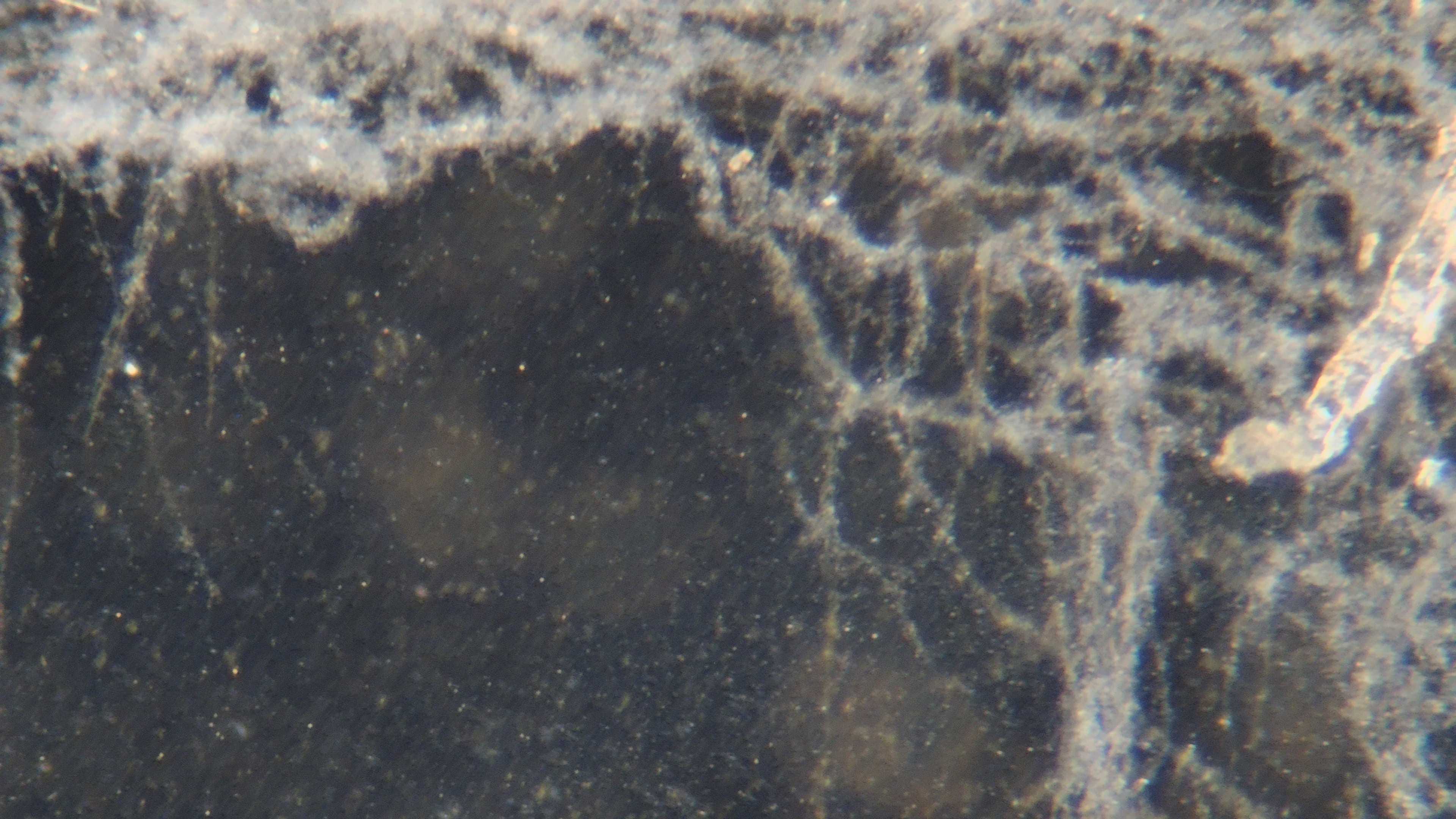
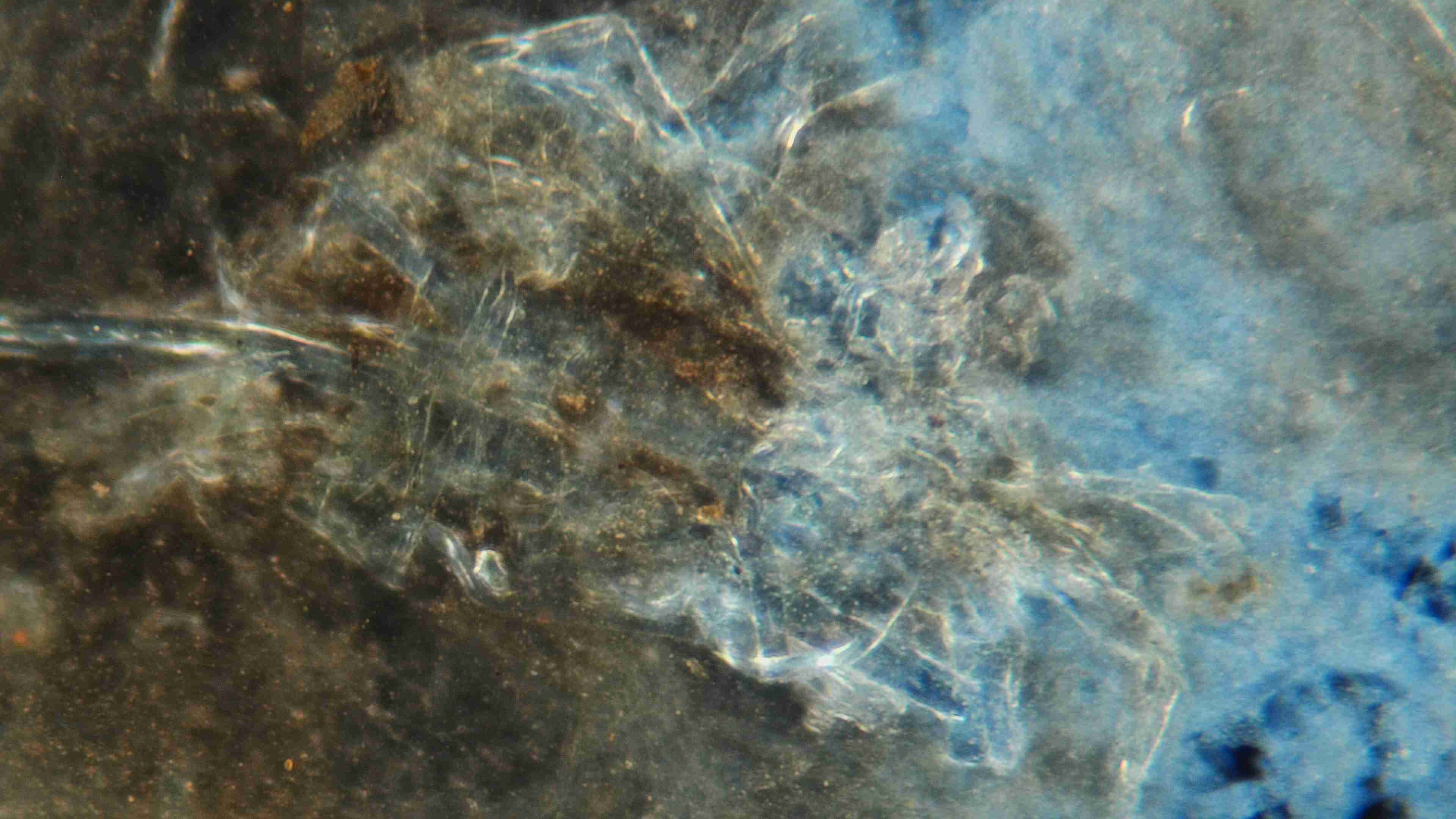
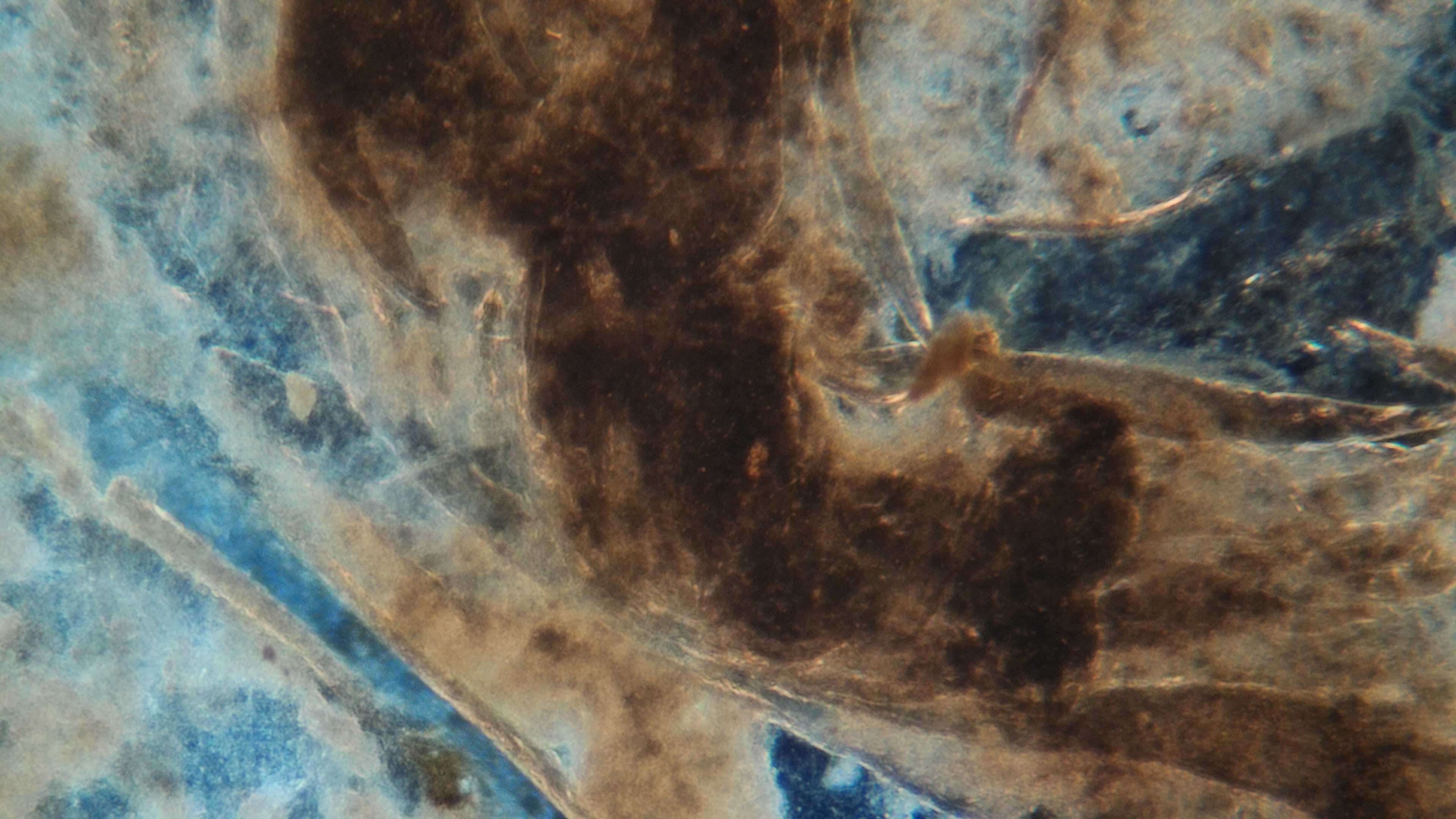
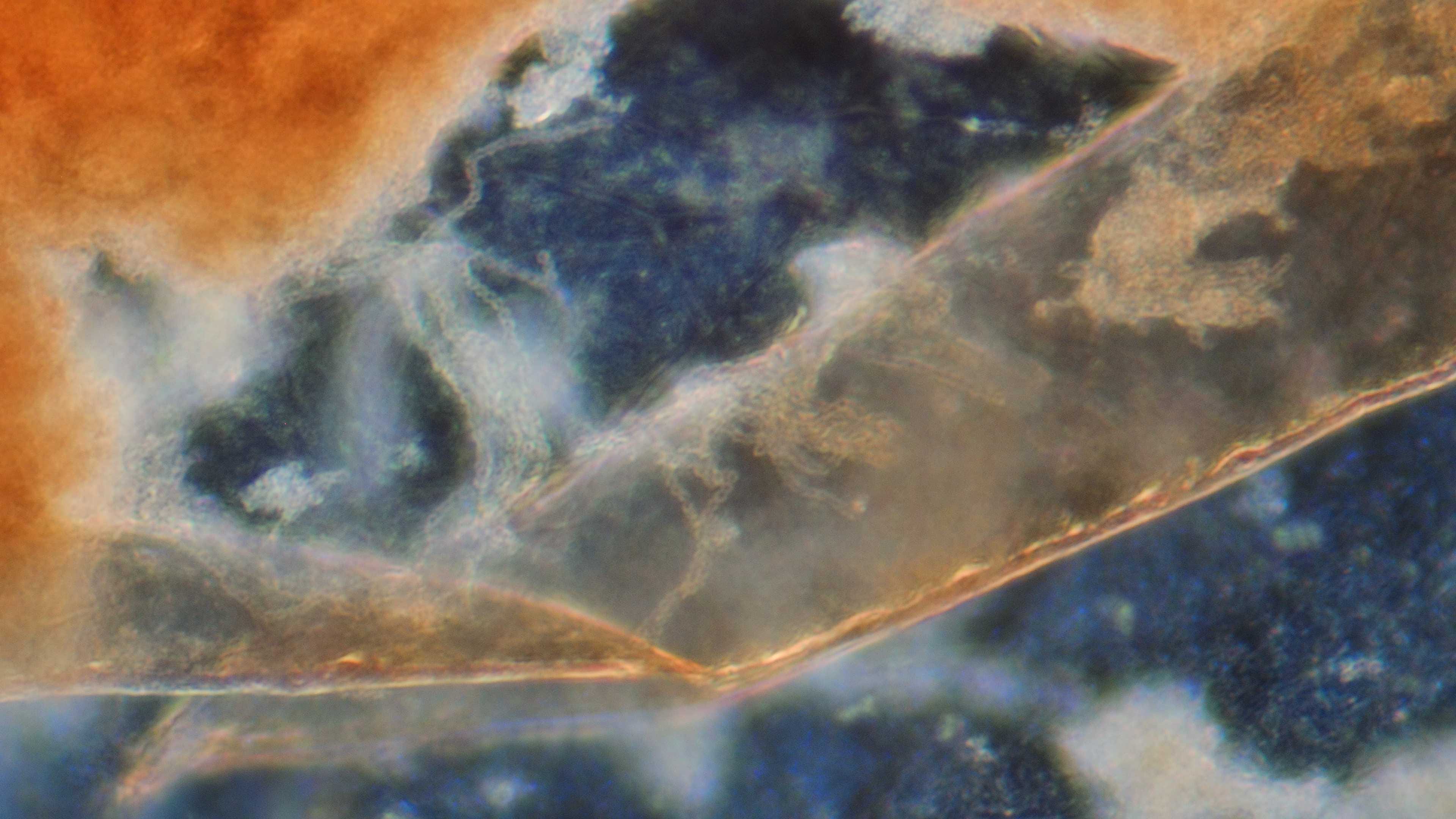
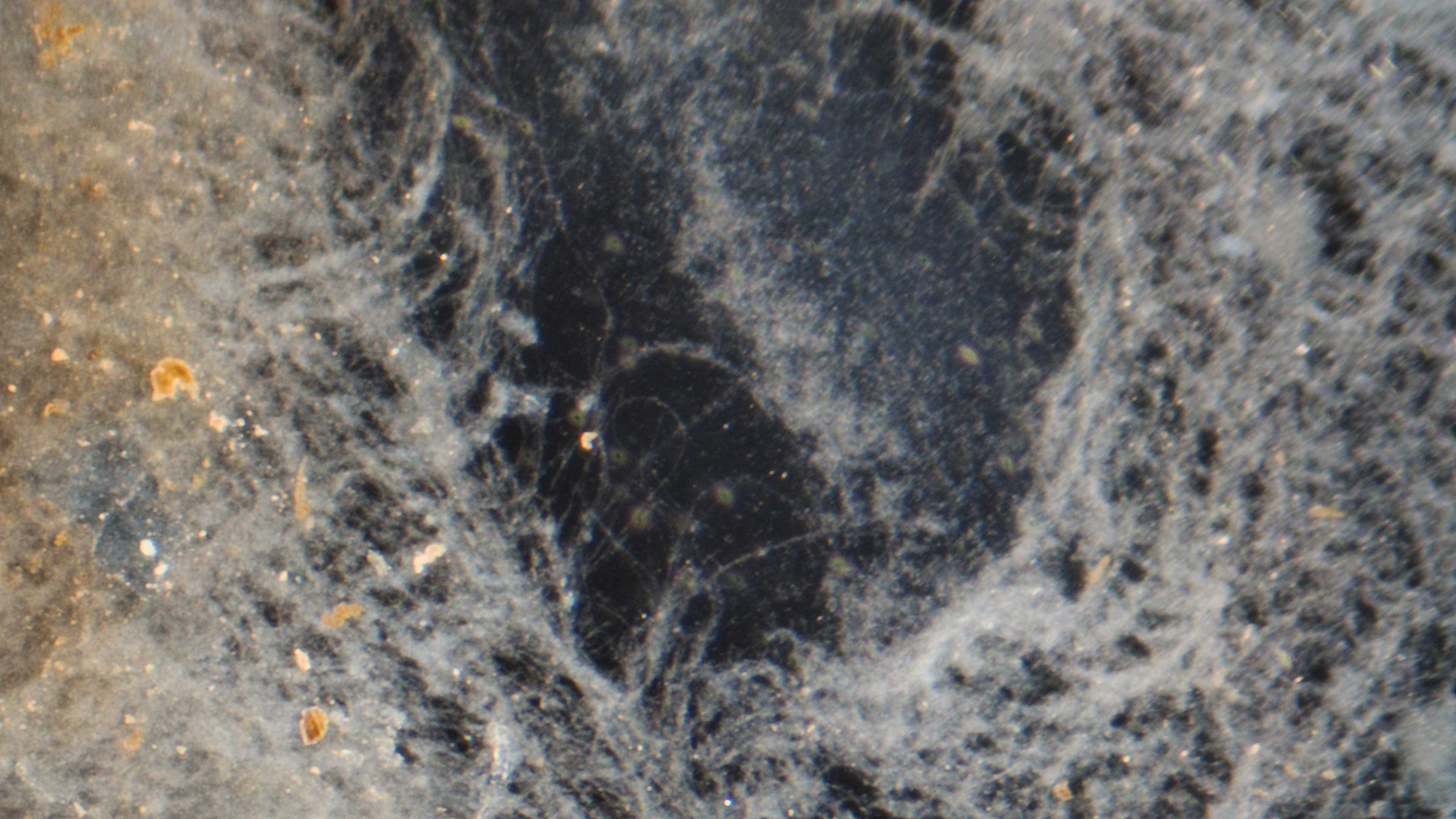
Again, I remind you that this was blood (not very much) and distilled water.


As stated, this appears to be a form of synthetic tissue scaffolding, which is being produced rapidly and in large quantities by the CDB in response to the application of electrical energy.
Video Footage
We shall now turn attention to a live video of this transformation process. None of the following videos is sped up or altered (except trimmed) in any way.
I apologize if there is erratic/frustrating motion; these are clips from longer extended views of this ‘reaction.’
This process was captured using the following microscope fixture:
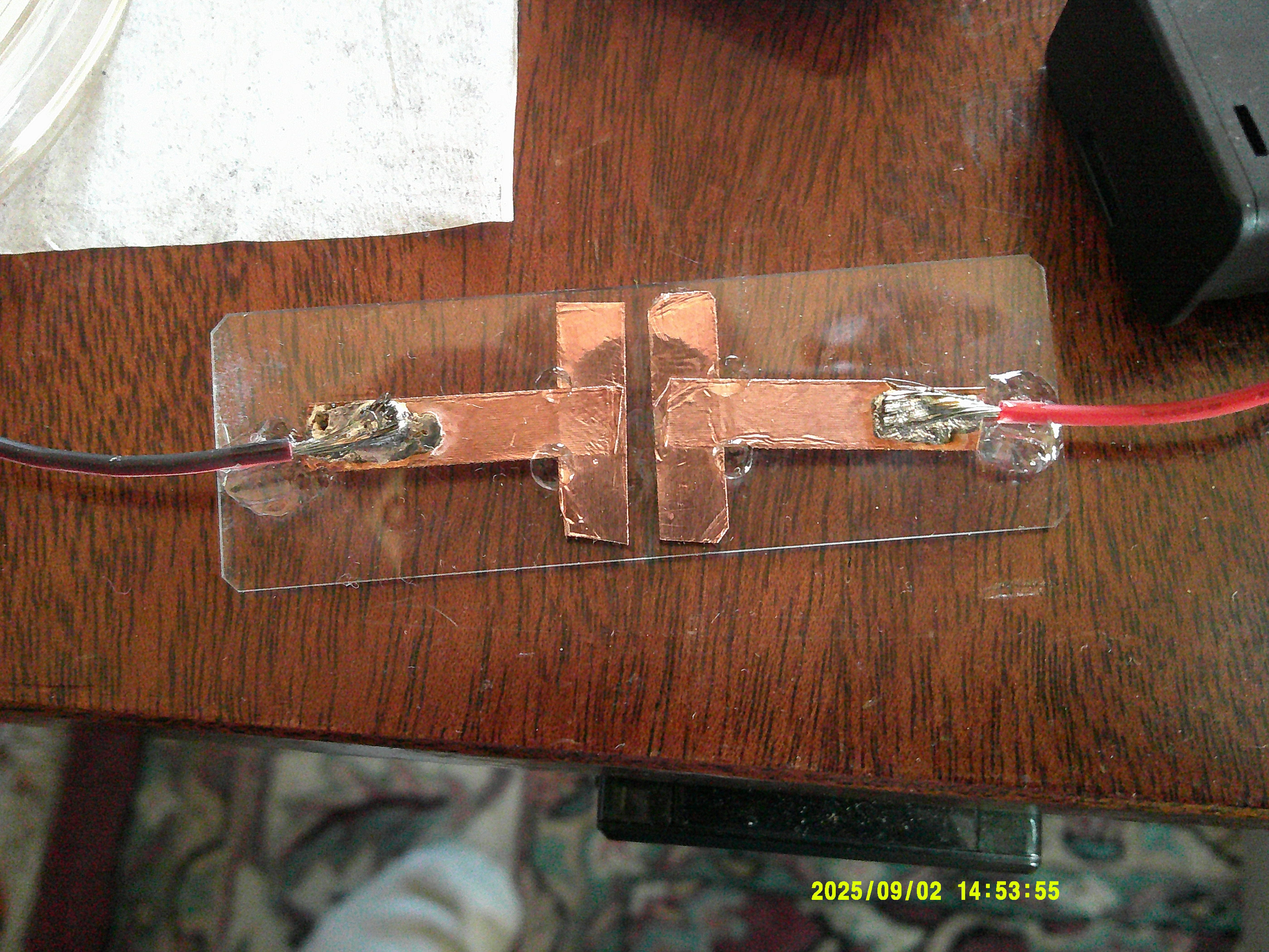
Deposition of sample so that it bridges the strips on either side allow for completion of electrical circuit, while being viewed under the microscope.
Videos are in brightfield. Voltage applied 2.4v. The mixture used was a ‘typical’ dilution (several mL distilled water and less than 5 drops of blood). Most video footage is 10x/20x objective.
There is clearly very complex structural assembly here.
Close up of CDBs actively constructing tissue scaffolding.
Truly remarkable organization and rapid formation.
Please note that the blood/water mixture in the following two videos was allowed to sit for several days; hence, the red blood cell membranes have mostly broken down. The important subject of synthetic erythrocytes (and other cells) will not be touched on at the moment.
The straight line forms are CDBs that have assembled themselves into a linear chain. Again, synthetic biology and related metabolic products absolutely dominate the picture.
5sec-15sec in the above video shows the obvious rapid formation of a large complex tissue filament-like structure.
Countless active CDBs can be seen in very large numbers throughout the sample.
Once again, I suggest the primary driver is the overwhelming presence of synthetic biology (CDB) and associated metabolic products, the production of which seems to be greatly accelerated with the application of small amounts of electrical energy.
There is extraordinarily advanced synthetic biological engineering (and other highly advanced nanotechnology/energetics) at play here.
The extreme involvement of electromagnetic energy sources is to be highlighted…brightly.
These claims are completely in line with decades of Carnicom Institute research, particularly in the paper referenced at the beginning of this article. The entire six part series should be read and understood by all.
I have many more images, videos, and insights/theories related to this process and hope to expand on them in a future post.
Thank you for taking the time to read this article!
Michael Merrick
Hi Michael,
Nice work. Was the growth off the + or _ terminal?
Cheers, m
Thank you for this amazing work!